Advancements in PET Imaging for Cancer Diagnosis
Introduction
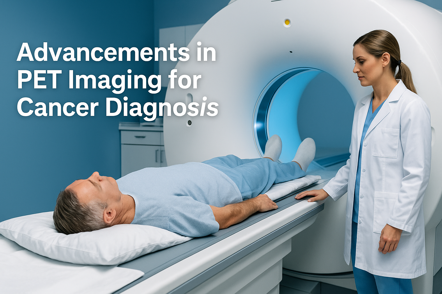
PET imaging for cancer diagnosis has become a cornerstone in modern oncology, offering unmatched precision in detecting, staging, and monitoring cancer. Positron Emission Tomography (PET) provides functional insights that traditional imaging cannot, helping clinicians make early, accurate decisions for patient care. With the latest technological advancements, PET scans are now faster, safer, and more informative, allowing for earlier detection and improved treatment outcomes.
What is PET Imaging?
PET (Positron Emission Tomography) uses a small amount of radioactive tracer to visualize metabolic processes in the body. Unlike CT or MRI, which focus on anatomy, PET highlights areas of abnormal activity, making it invaluable for detecting cancerous growths, evaluating tumor response, and monitoring recurrence.
Key Advancements in PET Imaging
-
Hybrid PET/CT and PET/MRI
Modern imaging often combines PET with CT or MRI, providing both functional and structural information. This hybrid approach allows clinicians to precisely locate tumors, assess their size, and understand their metabolic activity, which is essential for planning targeted therapies.
-
Advanced Radiotracers
The development of new radiotracers has expanded the ability of PET scans to detect specific cancer types. For instance, tracers targeting prostate or neuroendocrine tumors improve diagnostic accuracy and help in monitoring treatment response. Learn more about advanced PET tracers at RadiologyInfo – PET Scans.
-
Quantitative Imaging and AI Integration
Artificial intelligence (AI) is now being used to enhance PET image interpretation, reduce scan times, and improve tumor detection. AI-driven algorithms provide more precise quantification of tumor metabolism, enabling personalized treatment plans and better patient outcomes.
-
Reduced Radiation Dose and Faster Scans
Newer PET systems require smaller tracer doses while producing high-quality images. This advancement ensures patient safety while reducing time spent in the scanner, making PET scans more comfortable and accessible.
Benefits of PET Imaging for Cancer Diagnosis
- Early and accurate detection of cancer
- Differentiation between benign and malignant lesions
- Monitoring response to therapy
- Detection of cancer recurrence
- Guidance for targeted biopsies and radiotherapy
For patients seeking advanced cancer imaging, scans complement other diagnostic tools like CT and MRI, offering a comprehensive view of tumor biology. If you want to learn more about complementary imaging methods, visit Request an Appointment – Lake Zurich Open MRI.
Who Should Consider PET Imaging?
PET imaging is recommended for patients who:
- Have been diagnosed with cancer and require staging
- Need assessment of treatment response
- Are suspected to have cancer recurrence
- Require evaluation of unexplained symptoms related to cancer
By combining PET imaging with conventional imaging, clinicians can develop highly personalized treatment strategies that improve survival rates and quality of life.
Book Your Scan at Lake Zurich Open MRI
At Lake Zurich Open MRI, we provide state-of-the-art imaging with a patient-friendly environment, timely scheduling, and expert interpretation. Our team ensures your diagnostic journey is smooth, accurate, and compassionate.
Request your appointment today: Lake Zurich Open MRI Imaging Center

