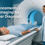
Diagnosing Cardiac Arrhythmias with MRI: A Breakthrough in Heart Health

Introduction
Cardiac arrhythmias are disorders of the heart’s rhythm that can range from harmless to life-threatening. Whether the heart beats too fast, too slow, or irregularly, diagnosing these conditions early is critical to preventing complications like stroke, heart failure, or cardiac arrest. Thanks to advancements in medical imaging, Magnetic Resonance Imaging (MRI) is now playing a pivotal role in accurately diagnosing arrhythmias.
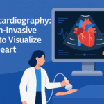
MRI offers a safe, non-invasive, and highly detailed way to visualize the heart’s structure and function, making it a powerful tool for patients and doctors alike. In this article, we will explore how MRI aids in diagnosing cardiac arrhythmias, its benefits, the procedure, and what patients should know.
What Are Cardiac Arrhythmias?
Cardiac arrhythmias occur when the electrical signals that control the heart’s rhythm become irregular. These can manifest as:
- Atrial Fibrillation (AFib): Irregular, rapid heartbeats.
- Bradycardia: Abnormally slow heart rate.
- Ventricular Tachycardia: Fast, abnormal rhythms originating from the heart’s ventricles.
- Premature Ventricular Contractions (PVCs): Extra heartbeats that disrupt normal rhythm.
While some arrhythmias may cause mild symptoms like palpitations or fatigue, others can increase the risk of severe complications, underscoring the importance of early and accurate diagnosis.
How MRI Helps in Diagnosing Cardiac Arrhythmias
1. Detailed Structural Imaging
MRI provides highly detailed images of the heart’s anatomy, enabling doctors to identify abnormalities such as:
- Enlarged heart chambers
- Damaged or scarred heart tissues
- Changes in heart wall thickness
These structural abnormalities often serve as triggers or underlying causes of arrhythmias.
2. Tissue Characterization
Specialized MRI techniques, like Late Gadolinium Enhancement (LGE), allow doctors to map scar tissue and fibrosis in the heart. Scar tissue disrupts the normal electrical pathways of the heart, leading to irregular rhythms. By pinpointing these areas, MRI helps doctors determine the exact source of the arrhythmia.
3. Real-Time Functional Analysis
Cardiac MRI not only captures static images but also evaluates the heart in motion. This helps assess:
- Blood flow through the heart and major vessels
- Ventricular function (how well the heart is pumping)
- Valve performance
Such dynamic assessments are crucial in diagnosing arrhythmias like ventricular tachycardia or atrial fibrillation.
Why Choose MRI Over Other Diagnostic Tools?
While traditional tools like ECGs, Holter monitors, and echocardiograms remain valuable, MRI offers unique advantages for diagnosing arrhythmias:
- Comprehensive Imaging: MRI evaluates both the heart’s structure and function, giving a complete picture in a single test.
- Non-Invasive and Radiation-Free: Unlike X-rays or CT scans, MRI uses no ionizing radiation, making it safer for repeated use, especially in younger patients.
- Highly Accurate Diagnosis: MRI’s ability to detect scar tissue and characterize heart function makes it a gold standard for diagnosing complex arrhythmias.
- Personalized Treatment Planning: The detailed information obtained from MRI allows doctors to tailor treatments, from medications to catheter ablations.
What to Expect During a Cardiac MRI
If your doctor has recommended an MRI for diagnosing cardiac arrhythmias, here’s what the procedure typically involves:
Before the MRI
- Preparation: Wear comfortable clothing and remove any metal objects, such as jewelry or watches.
- Medical History: Inform your doctor if you have metal implants (e.g., pacemakers or stents) or if you are pregnant.
- Fasting: You may be asked to avoid eating or drinking for a few hours before the scan.
During the MRI
- Positioning: You will lie on a flat table that slides into the MRI machine.
- Scan Duration: The procedure usually takes 30-60 minutes.
- Sounds: The machine makes loud tapping or thumping noises during the scan, so earplugs or headphones will be provided.
- Contrast Agent: In some cases, a contrast dye (like gadolinium) is injected to highlight areas of the heart. This is typically safe and enhances the imaging of scar tissue.
After the MRI
- No Recovery Time: You can resume normal activities immediately.
- Results: Your doctor will review the images and discuss the findings during a follow-up appointment.
Benefits of Using MRI for Arrhythmias
- Early and Accurate Diagnosis: MRI identifies subtle changes in heart tissue that other imaging techniques may miss.
- Better Treatment Outcomes: By pinpointing the cause of arrhythmias, MRI helps in planning targeted treatments like ablation therapy or medications.
- Safe for Most Patients: With no radiation exposure, MRI is suitable for most individuals, including those needing regular monitoring.
- Holistic Understanding: MRI provides a complete view of the heart’s structure and function, ensuring no detail is overlooked.
Are There Any Limitations?
While MRI is an excellent diagnostic tool, it does have some limitations:
- Not Suitable for All Patients: Individuals with certain metal implants, severe claustrophobia, or kidney issues (in case of contrast dye use) may not be candidates for MRI.
- Cost and Availability: MRI can be more expensive and less accessible compared to other imaging methods.
- Contrast Agent Risks: Though rare, allergic reactions to contrast agents can occur.
Conclusion
Diagnosing cardiac arrhythmias with MRI has transformed the way these heart conditions are detected and treated. By offering unparalleled imaging detail, tissue characterization, and functional analysis, MRI empowers healthcare providers to make accurate diagnoses and develop personalized treatment plans.
If you’re experiencing symptoms like palpitations, dizziness, or chest discomfort, speak with your cardiologist about whether an MRI might be right for you. Early detection through advanced tools like MRI can save lives and improve quality of life.
With its non-invasive nature and radiation-free technology, cardiac MRI is paving the way for safer and more effective heart care, ensuring better outcomes for patients around the world.
If you need your scans done please call our number (847) 726-0674 or visit our Request an Appointment page.

