Radiology in Rheumatology: Imaging Arthritis and Autoimmune Diseases

Introduction: Radiology in Rheumatology – A Powerful Tool for Diagnosing Arthritis and Autoimmune Diseases
Arthritis and autoimmune diseases can cause chronic pain, inflammation, and debilitating symptoms. Radiology in rheumatology plays a vital role in diagnosing these conditions early, helping physicians assess joint damage, inflammation, and disease progression. Imaging techniques like X-ray, MRI, and ultrasound are indispensable tools for rheumatologists to identify arthritis and autoimmune diseases such as rheumatoid arthritis (RA), lupus, and ankylosing spondylitis.
In this article, we’ll explore how radiology in rheumatology aids in diagnosing these conditions and monitoring treatment effectiveness.
Why Radiology is Crucial in Rheumatology
Radiology in Rheumatology: Imaging Arthritis and Autoimmune Diseases with Precision
Rheumatic conditions can be challenging to diagnose because symptoms often overlap with other disorders. Early and accurate imaging is key to:
✔ Detecting joint inflammation, synovitis, and early signs of joint damage
✔ Identifying bone erosions, tendonitis, and cartilage damage
✔ Monitoring disease progression and treatment response
✔ Assessing the impact of autoimmune diseases on organs and soft tissues
Related Reading: Common Rheumatic Conditions Diagnosed with Imaging
Key Imaging Techniques in Rheumatology
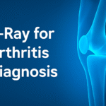
1. X-Ray Imaging for Arthritis
-
First-line imaging for assessing joint damage in arthritis
-
Identifies bone spurs, joint space narrowing, and erosions
-
Most commonly used for osteoarthritis and rheumatoid arthritis
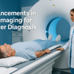
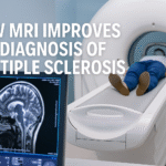
2. MRI for Joint and Soft Tissue Imaging
-
Provides high-resolution images of soft tissues and cartilage
-
Ideal for diagnosing rheumatoid arthritis, ankylosing spondylitis, and lupus
-
MRI is used to detect synovial inflammation and bone marrow edema
-
Offers better sensitivity for detecting early disease compared to X-ray
3. Ultrasound for Musculoskeletal Imaging
-
Real-time imaging to assess joint fluid, bursitis, and tendonitis
-
Used to guide injections or biopsies
-
Highly effective in monitoring synovitis and joint inflammation in rheumatoid arthritis
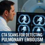
4. CT Scans for Bone and Joint Evaluation
-
Helpful for detecting bone damage, especially in complicated fractures or advanced rheumatic disease
-
Provides detailed 3D views of joints and bones
Conditions Diagnosed with Imaging in Rheumatology
✔ Rheumatoid arthritis – Joint inflammation, bone erosion, and soft tissue damage
✔ Osteoarthritis – Degenerative joint disease causing cartilage loss and bone changes
✔ Ankylosing spondylitis – Inflammation of the spine and sacroiliac joints
✔ Psoriatic arthritis – Inflammation affecting the skin and joints
✔ Systemic lupus erythematosus (SLE) – Autoimmune disease affecting joints, skin, and internal organs
✔ Gout – Build-up of uric acid crystals in the joints
✔ Sjögren’s syndrome – Dry eyes, mouth, and joint inflammation
Need Rheumatology Imaging? Book Your Appointment Today
Benefits of Radiology in Rheumatology
✔ Early Detection – Identifies early joint changes before symptoms become severe
✔ Accurate Diagnosis – Helps differentiate between inflammatory and degenerative arthritis
✔ Guides Treatment Decisions – Helps doctors decide between medication, physical therapy, and surgical options
✔ Monitors Disease Progression – Tracks the impact of treatment and joint health over time
✔ Minimizes Invasive Procedures – Non-invasive imaging reduces the need for biopsies or exploratory surgeries
What to Expect During a Rheumatology Imaging Procedure
Radiology in Rheumatology: Imaging Arthritis and Autoimmune Diseases – Patient Experience
1. Before the Scan
✔ Minimal preparation is required for most imaging tests
✔ MRI or ultrasound may require you to avoid eating or drinking for several hours
✔ Inform your doctor of any implants, allergies, or medical conditions (for contrast agents)
2. During the Scan
-
For X-ray, you will be positioned near the machine to capture the required joint images
-
For MRI, you will lie on a table while the machine captures detailed images of your joints or soft tissues
-
For ultrasound, a gel is applied to the skin to visualize the joint in real time
3. After the Scan
✔ Most patients resume normal activities immediately
✔ A radiologist reviews the images and sends a report to your rheumatologist
✔ Your doctor will discuss the results and next steps, whether that’s medication, physical therapy, or further tests
Related Topic: How to Prepare for Rheumatology Imaging
Conclusion
Radiology in rheumatology is a game-changer for diagnosing arthritis and autoimmune diseases. With X-rays, MRI, and ultrasound, doctors can detect joint inflammation, bone damage, and disease progression at earlier stages, enabling more effective treatments and better outcomes for patients.
If you’re experiencing symptoms of arthritis or autoimmune conditions, ask your doctor if imaging can help clarify your diagnosis.
Need Imaging for Arthritis or Autoimmune Disease? Book Your Appointment Today


