Introduction
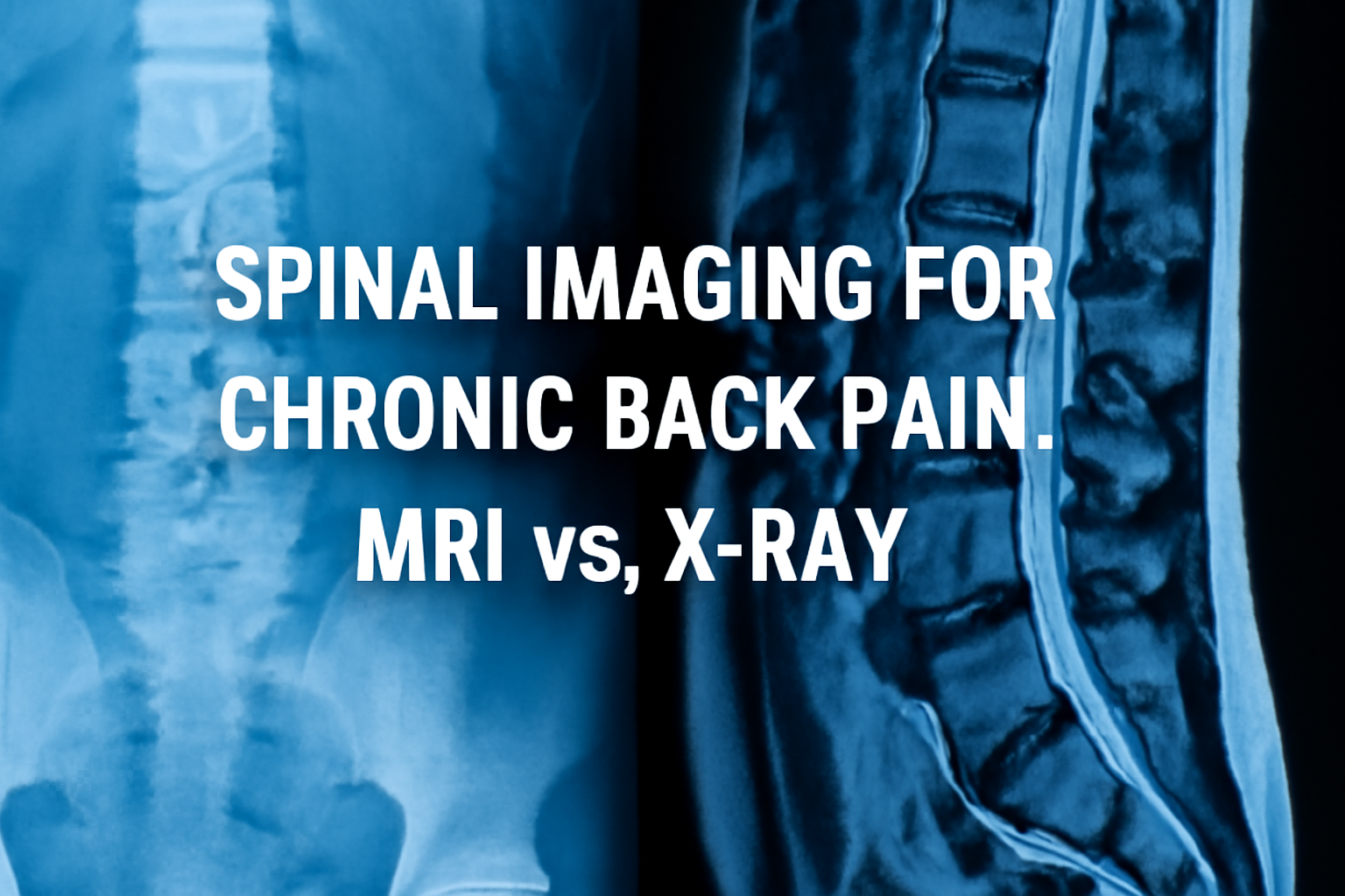
Spinal imaging for chronic back pain is a crucial part of diagnosing the underlying cause of persistent back pain. With a variety of imaging techniques available, two of the most common are MRI and X-ray. Both have their advantages, but understanding their differences is key to choosing the best imaging method for accurate diagnosis and effective treatment. In this article, we’ll explore the roles of MRI and X-ray in assessing chronic back pain and how each technique is used in clinical practice.
What is MRI for Spinal Imaging?
Magnetic Resonance Imaging (MRI) is an imaging technique that uses powerful magnets and radio waves to create detailed images of soft tissues, such as muscles, ligaments, discs, and nerves. Unlike X-rays, MRI doesn’t use ionizing radiation, which makes it a safer option, especially for individuals requiring frequent scans.
MRI for spinal imaging can reveal:
- Herniated discs
- Nerve compression
- Spinal stenosis
- Tumors or infections
For a deeper dive into how MRI works for spinal conditions, check out RadiologyInfo’s overview of MRI.
The Role of X-Ray in Spinal Imaging for Chronic Back Pain
X-ray imaging uses radiation to capture images of bones and joints, making it an excellent tool for identifying bone fractures, joint misalignments, and degenerative changes such as osteoarthritis. While X-rays do not provide detailed soft tissue images, they can effectively detect bone-related issues that contribute to chronic back pain.
X-ray is commonly used to diagnose:
- Fractures or misalignment
- Scoliosis
- Spinal degeneration
Although X-ray is often the first choice for evaluating bone abnormalities, it has limitations in visualizing soft tissues or nerve damage. However, for an initial evaluation, X-ray remains a valuable tool.
MRI vs. X-Ray: When to Use Each for Spinal Imaging
When deciding between MRI and X-ray for spinal imaging for chronic back pain, the choice largely depends on the clinical presentation:
When MRI is Preferred:
- If the patient has radiating pain down the legs or numbness.
- To assess soft tissue abnormalities, such as disc herniation or spinal nerve impingement.
- In cases where surgical planning is needed.
When X-Ray is Sufficient:
- For detecting bone fractures or spinal misalignments.
- In cases where only bone issues are suspected, such as early-stage osteoarthritis.
- As a preliminary step before considering more advanced imaging like MRI.
While MRI provides superior soft tissue detail, X-rays are cost-effective and faster, making them useful for quick initial assessments.
Advantages and Disadvantages of MRI and X-Ray for Chronic Back Pain
Advantages of MRI for Spinal Imaging:
- Provides detailed soft tissue images.
- No radiation exposure.
- Essential for nerve evaluation.
Disadvantages of MRI:
- More expensive than X-ray.
- Longer procedure time.
- Not suitable for patients with metal implants or claustrophobia.
Advantages of X-Ray for Spinal Imaging:
- Quick and cost-effective.
- Best for bone-related issues like fractures or alignment problems.
Disadvantages of X-Ray:
- Cannot visualize soft tissues or nerves.
- Uses radiation, though in small doses.
Conclusion
Spinal imaging for chronic back pain is essential for proper diagnosis and treatment. MRI is the preferred option when assessing soft tissues, nerves, or spinal cord issues, while X-ray remains an invaluable tool for evaluating bone abnormalities. The combination of both can often provide a comprehensive picture of the patient’s condition, leading to the most effective treatment.
If you’re experiencing chronic back pain and need detailed diagnostic imaging, schedule an appointment with Lake Zurich Open MRI for the best imaging care.


